
1. Introduction
Human Immunodeficiency Virus type 1 (HIV-1), the causative agent of AIDS, belongs to the Lentivirinae subfamily of retroviruses, a group of viruses characterized by slow progression of disease and long latency periods before clinical manifestations appear. The term lentivirus was originally introduced in 1954 by Sigurdsson to describe slowly progressive infectious diseases observed in sheep.
Lentiviruses include several animal and human pathogens such as HIV-1, HIV-2, simian immunodeficiency virus, feline immunodeficiency virus, equine infectious anemia virus, and others. A defining feature of this viral family is their dual tropism:
- They infect immune system cells, particularly monocytes and macrophages
- They exhibit neurotropism, meaning they can invade and persist in the central nervous system (CNS)
This dual targeting explains why lentiviral infections are both immunological and neurological diseases, with symptoms developing after prolonged periods of latency ranging from months to years.
Clinically, lentiviral infections can lead to:
- Immunodeficiency syndromes (as in HIV infection)
- Chronic inflammatory conditions
- Neurological disorders such as encephalitis
- Multisystem damage including hematologic and renal complications
The severity and presentation of disease vary depending on both the viral strain and the host immune response.
2. Genomic Organization and Replication Cycle of HIV-1
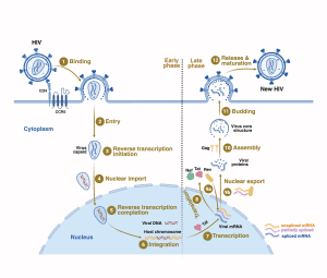
HIV-1 shares the basic genomic structure of retroviruses, encoding three essential gene groups:
- gag (structural proteins)
- pol (enzymes including reverse transcriptase)
- env (envelope glycoproteins)
However, HIV-1 is more genetically complex than most retroviruses because it also encodes several regulatory and accessory proteins, including:
- Tat
- Rev
- Nef
- Vif
- Vpu
- Vpr
2.1 Early Infection and Integration
After entry into a host cell, HIV-1 RNA is converted into DNA by reverse transcriptase. This viral DNA is then transported into the nucleus and integrated into the host genome, forming a provirus, allowing long-term persistence.
2.2 Gene Expression Regulation
Viral replication follows a tightly regulated cascade:
- Early phase transcription produces multiply spliced RNA encoding Tat, Rev, and Nef
- Tat protein amplifies viral transcription, enhancing viral RNA production
- Rev protein regulates RNA processing, shifting expression from early regulatory proteins to structural proteins
- This switch enables production of:
- Gag and Pol proteins (from unspliced RNA)
- Vif, Vpr, Vpu (from singly spliced RNA)
2.3 Functional Roles of Accessory Proteins
- Vif and Vpu enhance viral particle maturation and release
- Vpu promotes degradation of CD4 receptors, facilitating viral assembly
- Vpr plays roles in nuclear transport and viral replication efficiency
- Nef modulates immune evasion and downregulates CD4 expression; it may also enhance viral replication in vivo
3. Cellular Regulation and Viral Tropism
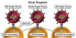
HIV-1 gene expression is strongly influenced by host transcription factors binding to the viral long terminal repeat (LTR), including:
- NF-κB
- SP1
- CTF/NF-1
- LBP-1
- EBP-1
Among these, NF-κB is particularly important, as it can reactivate latent virus during immune activation or inflammation (e.g., TNF-α or IL-1 stimulation).
A key biological characteristic of HIV-1 is its ability to replicate in:
- Activated T lymphocytes
- Terminally differentiated macrophages
Unlike many viruses, HIV-1 does not require actively dividing cells, which contributes to its persistence in long-lived cellular reservoirs.
4. HIV-1 Entry into the Central Nervous System
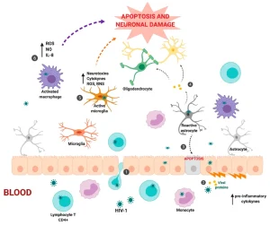
HIV-1 can invade the CNS very early after systemic infection, sometimes within weeks. Entry may occur without obvious neurological symptoms, although in some cases it manifests as acute meningitis or meningoencephalitis.
4.1 Acute Neurological Phase
Acute CNS infection typically occurs 3–6 weeks after primary infection and may include:
- Headache and fever
- Meningeal irritation
- Seizures
- Cognitive disturbances
- Flu-like systemic symptoms resembling infectious mononucleosis
CSF findings often include:
- Mild protein elevation
- Lymphocytic pleocytosis
- Normal glucose levels
During this stage:
- Serological tests may still be negative
- Viral antigen (p24) or RNA may be detectable
- Diagnosis often requires PCR or viral culture
In most cases, this early CNS invasion is clinically silent and underdiagnosed.
5. HIV-1–Associated Neurocognitive Disorders
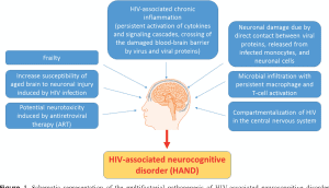
One of the most significant neurological consequences of HIV infection is HIV-1-associated cognitive-motor complex, also known as HIV encephalopathy or AIDS dementia complex.
5.1 Epidemiology and Frequency
The prevalence varies depending on disease stage and treatment era:
- Occurs in a significant proportion of advanced AIDS cases
- Can appear in 7–17% of patients depending on cohort
- May be present in subclinical form in a much larger fraction
Antiretroviral therapy, especially early regimens such as zidovudine, has significantly reduced incidence but has not eliminated it.
6. Clinical Features of HIV-1 Encephalopathy
The disorder typically develops gradually and insidiously, with progressive neurological decline.
6.1 Cognitive Symptoms
- Memory impairment
- Difficulty concentrating
- Slowed thinking (bradyphrenia)
- Language disturbances
- Executive dysfunction
- Social withdrawal
6.2 Motor Symptoms
- Gait disturbances
- Fine motor impairment
- Tremor
- Bradykinesia
- Muscle weakness
- Loss of coordination
Advanced disease may resemble Parkinsonian syndromes, including rigidity and postural instability.
6.3 Psychiatric Manifestations
- Depression (often resistant to treatment)
- Psychosis in some cases
- Mania or mood instability
- Delirium with hallucinations
- Cognitive-affective disorders
Importantly, psychiatric symptoms may be mistaken for primary psychiatric disease, delaying diagnosis.
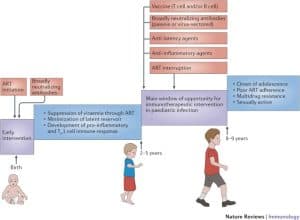
7. Pediatric HIV-1 Neuroinfection
In children, HIV-1 infection of the CNS often leads to:
- Developmental delay
- Loss of previously acquired milestones
- Spasticity and motor dysfunction
- Microcephaly
- Seizures and myoclonus
Neurological deterioration frequently parallels immune system decline, although it may also appear early in life.
8. Neuropathology of HIV-1 Brain Infection
Postmortem studies reveal characteristic structural changes:
8.1 Macroscopic Findings
- Brain atrophy
- Ventricular enlargement
- Cortical thinning
8.2 Microscopic Features
- White matter damage and demyelination
- Microglial nodules
- Astrocytosis
- Perivascular inflammation
- Multinucleated giant cells (hallmark feature)
- Synaptic and dendritic loss
- Neuronal reduction in cortical and subcortical regions
The basal ganglia are frequently affected, explaining motor symptoms and sensitivity to dopamine-blocking drugs.
Importantly, severity of pathology does not always correlate with clinical symptoms, indicating complex indirect mechanisms of injury.
9. Neurotropism: How HIV-1 Reaches Brain Cells
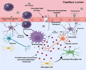
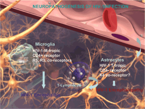
HIV-1 does not primarily infect neurons. Instead, CNS infection involves:
- Microglial cells (resident macrophage-like cells)
- Infiltrating monocytes/macrophages
- Occasionally astrocytes
Evidence of viral presence has been demonstrated using:
- In situ hybridization
- Immunocytochemistry
- PCR-based detection
- Electron microscopy
Microglia and macrophages act as major viral reservoirs within the brain, supporting persistent infection.
10. Mechanisms of Brain Injury in HIV-1 Infection
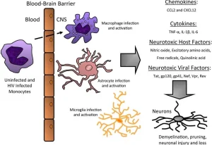
Neuronal damage in HIV is largely indirect rather than direct, involving multiple pathways:
10.1 Viral Protein Toxicity
- gp120 can induce neuronal calcium dysregulation and excitotoxicity
- Tat protein can alter membrane function and neuronal signaling
- Nef protein may influence ion channel activity and immune evasion
10.2 Excitotoxicity and Calcium Dysregulation
- Overactivation of NMDA receptors
- Excess glutamate signaling
- Calcium overload leading to neuronal apoptosis
10.3 Cytokine-Mediated Injury
Activated microglia and macrophages release:
- TNF-α
- IL-1β
- IL-6
These cytokines contribute to:
- Blood–brain barrier disruption
- Myelin damage
- Astrocyte activation
- Neuroinflammation
10.4 Oxidative Stress and Metabolic Injury
Inflammatory mediators increase:
- Reactive oxygen species
- Mitochondrial dysfunction
- Synaptic degeneration
11. Conclusion
HIV-1 infection of the brain represents a complex neuroimmunological disorder driven by a combination of:
- Persistent viral reservoirs in microglia and macrophages
- Indirect neuronal injury mediated by viral proteins
- Chronic inflammatory cytokine production
- Synaptic and white matter degeneration
Rather than being a purely infectious encephalitis, HIV-1 brain disease is best understood as a chronic neuroinflammatory and neurodegenerative process, influenced by both viral biology and host immune responses.
Advances in antiretroviral therapy have significantly reduced severe neurological complications, but subclinical cognitive impairment and long-term neuroinflammation remain important clinical challenges.